

Why in news?
Researchers at the Indian Institute of Technology Mandi developed a spiky ceramic coating for three-dimensional printed bone scaffolds. The surface resembles a sea urchin at the nanoscale. Its tiny projections physically damaged bacteria while supporting bone-forming activity in laboratory and animal tests.
The coating uses hydroxyapatite on a polylactic acid scaffold. The team reported effects against Escherichia coli and Staphylococcus aureus. Mouse experiments supported the antibacterial and bone-integration findings. This remains preclinical biomaterials research. It is not yet an approved implant treatment for patients. The research addresses infection and weak integration with surrounding bone. Further studies must test both functions.
The problem with bone implants
Large bone defects can follow trauma, infection, cancer surgery or congenital disease. A graft or implant must fill the defect and bear suitable loads. It should also help new bone attach and grow. Patient-specific three-dimensional printing can match complex shapes. Yet the printed material may remain biologically inactive without surface modification.
Infection creates a second difficulty. Bacteria can attach to an implant and form a biofilm. This protective community resists immune attack and many antibiotics. Infection can delay healing or force implant removal. Repeated antibiotic use also adds resistance pressure. A surface that discourages bacteria while welcoming bone cells would address both problems.
What the researchers built
The base scaffold uses polylactic acid, or PLA, a printable biodegradable polymer. PLA provides shape and structural support. Its surface is normally hydrophobic and relatively bioinert. The team first treated it with alkali. This created negatively charged chemical sites. Those sites helped mineral particles begin forming directly on the printed surface.
A low-temperature hydrothermal process then deposited hydroxyapatite. This calcium-phosphate mineral resembles an important mineral component of bone. It grew into clustered radial nanospicules. The resulting surface looked like many microscopic sea urchins. It also became rough, porous and highly water-attracting. These properties can affect both bacteria and mammalian cells.
How the antibacterial action differs
Many implant coatings release antibiotics, silver or other chemical agents. This design seeks a mechanical effect from surface topography. Sharp nanospicules can stress or puncture bacterial membranes after contact. The researchers call this mechano-bactericidal activity. It may reduce dependence on continuously released drugs. The coating still requires careful compatibility testing with healthy cells.
The study tested two common bacterial models. E. coli represents Gram-negative bacteria, while S. aureus is Gram-positive. Both can be relevant to medical infection, although real implant infections involve many strains. Laboratory results showed bacterial damage on the coated surface. A mouse infection model provided supporting evidence inside living tissue.
How the coating may support bone
Hydroxyapatite offers chemical signals familiar to bone tissue. Its rough and wettable surface can improve cell attachment. The team tested MG-63 cells, a widely used bone-related cell line. Coated scaffolds supported collagen and calcium deposition. Subcutaneous implantation in mice also showed mineral formation. These findings suggest osteointegration potential rather than completed bone repair.
Osteointegration means stable direct contact between living bone and an implant. It requires more than early cell attachment. Blood vessels must enter the repair area. New bone must remodel under mechanical load. Inflammation must stay controlled. Future studies need load-bearing defect models and longer follow-up. Human bone healing is more complex than an ectopic mouse implant.
What nature contributed
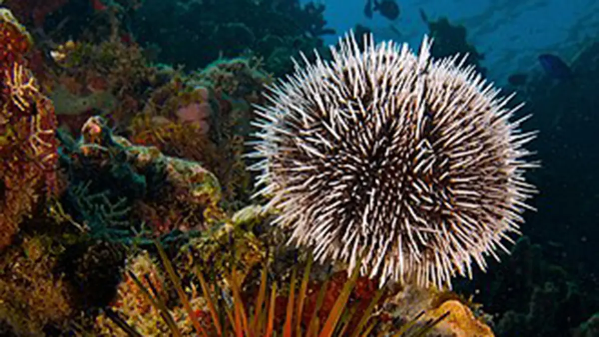
Sea urchins belong to class Echinoidea within phylum Echinodermata. They live only in marine environments and occur across the world’s oceans. A hard test of interlocking calcium-carbonate plates supports the body. Movable spines cover that test. Tube feet use a water vascular system for movement, feeding and sensing.
Many species graze algae using a jaw apparatus called Aristotle’s lantern. The apparatus contains five teeth and supporting structures. Sea urchins can maintain ecological balance by controlling algae. Excessive populations can also overgraze kelp and create barren reefs. Their role therefore depends on predators, food and habitat. The implant research borrows shape, not the animal’s whole biology.
Biomimicry needs accurate translation
Biomimicry studies a useful natural feature and adapts its principle. Here, the useful feature is radial surface roughness. The implant does not contain sea-urchin tissue. It also does not copy a living immune system. Engineers used hydroxyapatite to create a comparable nanoscale form. That distinction prevents an attractive metaphor from replacing the material science.
The same topography can affect different cell types differently. Bacteria are smaller and have distinct membranes. Mammalian cells can spread across larger surface features. However, a design that damages bacteria could still trigger inflammation or cell injury. Researchers must define safe dimensions and coating strength. Detached particles would create another safety concern.
Next steps towards clinical use
The team must test sterilisation, storage and large-batch manufacturing. Coatings should remain attached during surgery and loading. Degradation products need toxicity assessment. Animal studies should use realistic bone defects and compare standard implants. Researchers must also test more bacterial strains and mature biofilms. Regulatory evidence must cover both material and manufacturing process.
Cost will affect access. Low-temperature processing may reduce energy needs, but quality control can remain demanding. Printed implants also require good imaging and surgical planning. A successful coating would form one part of a treatment system. Infection control still includes sterile surgery, patient care and appropriate antibiotics. No surface can replace those measures alone.
Preclinical, not patient-ready
The coating showed useful effects in cell tests and mice. These findings support further development. They do not prove safe use in human bone. Load-bearing studies, manufacturing validation and regulatory review remain necessary.
Conclusion
The IIT Mandi research joins two important functions within one surface. The nanospicules aim to damage bacteria without releasing a conventional antibiotic. Hydroxyapatite simultaneously creates a more bone-friendly interface. That combination is scientifically attractive. Its value will depend on durable safety and performance in realistic bone-repair models.
The project also shows responsible use of natural inspiration. Scientists copied a structural idea instead of making vague claims about nature. Clear preclinical language is equally important. Further evidence may support patient-specific, infection-resistant implants. Until then, this remains a promising platform for biomaterials research.